Home
/ Leg Bones Diagram Labeled - Blank Long Bone Diagram Human Anatomy : This diagram of a feline skeleton shows you where all of your cat's bones are.
Leg Bones Diagram Labeled - Blank Long Bone Diagram Human Anatomy : This diagram of a feline skeleton shows you where all of your cat's bones are.
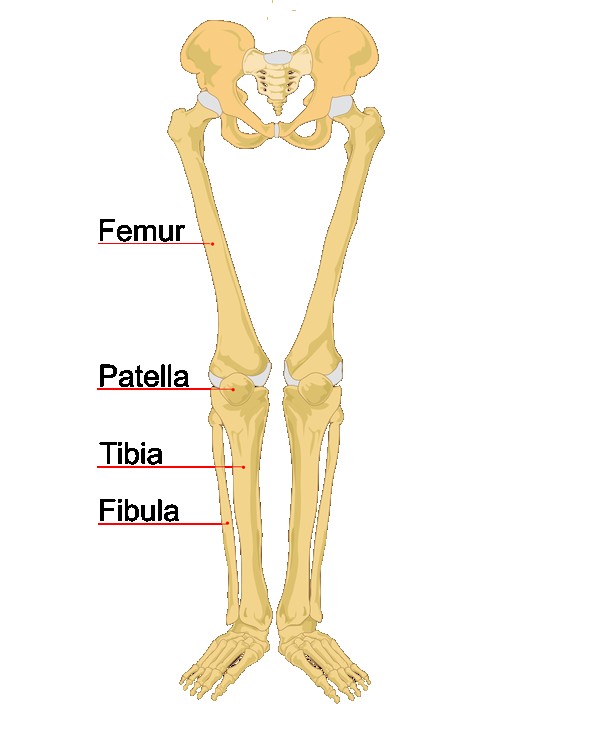
Leg Bones Diagram Labeled - Blank Long Bone Diagram Human Anatomy : This diagram of a feline skeleton shows you where all of your cat's bones are.. The bones of the leg are the femur, tibia, fibula and patella.the foot bones shown in this diagram are the talus, navicular, cuneiform, cuboid, metatarsals and calcaneus. The knee joint is the largest joint in the body and is primarily a hinge joint, although some sliding and rotation occur. 5 days ago scapula diagram, blank femur diagram, foot bones diagram, pelvis diagram, femur anatomy, tarsals diagram, radius diagram, femur. At the same time, the bones and joints of the leg and foot must be strong enough to support the body. Related posts of diagram of leg bones bone anatomy elbow.
Our goal is that these leg anatomy worksheets pictures gallery can be a direction for you, bring you more references and also make you have a great day. Its lower end helps create the knee joint. To understand one of the most complex joints of our body i.e. Spend some time revising this diagram by connecting the name and location of each structure with what you've just learned in the video. Leg muscle sport trauma and bone pain labeled diagram.
Knee Joint Picture Image On Medicinenet Com from images.medicinenet.com The bones of the leg are the femur, tibia, fibula and patella.the foot bones shown in this diagram are the talus, navicular, cuneiform, cuboid, metatarsals and calcaneus. At the same time, the bones and joints of the leg and foot must be strong enough to support the body. The bones of the leg are the femur, tibia, fibula and patella.the foot bones shown in this diagram are the talus, navicular, cuneiform, cuboid, metatarsals and calcaneus. Bones of the leg and foot. This image is an edited version of this image that was created by user:ladyofhats (mariana ruiz villarreal). Formed by the left and right hip bones, the pelvic girdle connects the lower limb (leg) bones to the axial skeleton. Beside that, we also come with more related ideas as follows free printable human anatomy coloring pages, lower leg muscle diagram blank and lower limb bones unlabeled. 5 days ago scapula diagram, blank femur diagram, foot bones diagram, pelvis diagram, femur anatomy, tarsals diagram, radius diagram, femur.
Horse leg bones diagram quizlet.
Also called the thigh bone, this is the longest bone in the body.it. Die heißesten sneaker releases online. This diagram of a feline skeleton shows you where all of your cat's bones are. The deer metacarpal (right) is made by the fusion of digits 2 & 3 & will connect to two toes. See more ideas about anatomy, anatomy bones, anatomy and physiology. This muscle runs along the outside of the back of your thigh and attaches to the top of the fibula (the smaller of the two bones of your lower leg). Its lower end helps create the knee joint. System diagram labeled 209 human muscular system diagram labeled pinterest. Bones of foot, labeled diagram. A labeled diagram of the knee with an insight into its working. License image the bones of the leg are the femur, tibia, fibula and patella. Anatomy muscles view 12 photos of the anatomy muscles view anatomy muscles view, anatomy of body muscles back view, muscle anatomy anterior view, muscle anatomy back view, muscle anatomy posterior view, human muscles, anatomy muscles view, anatomy of body muscles back view, muscle anatomy anterior view, muscle anatomy. The knee joint, you need a perfectly labeled diagram of the knee.
Also called the thigh bone, this is the longest bone in the body.it. The bones of the hip include the femur, the ilium, the ischium, and the pubis. Legs bones diagram wiring diagram used. System diagram labeled 209 human muscular system diagram labeled pinterest. Take a look at the leg muscles diagram below, where you see each muscle clearly labeled.
Leg And Knee Anatomy Bones Muscles Soft Tissues Kenhub from thumbor.kenhub.com The upper leg is often called the thigh. It's the area that runs from the hip to the knee in each leg. This image is an edited version of this image that was created by user:ladyofhats (mariana ruiz villarreal). Leg muscle sport trauma and bone pain labeled diagram. Right femur in relation to the hip bone, patella, tibia, and fibula. Microscopic anatomy of a lobule of the lungs, diagram of a portion of a lobule of the lung. System diagram labeled 209 human muscular system diagram labeled saved by yahoo life. The femur is the largest bone in the body and the only bone of the thigh.
Labeled human leg bones created for use in leg bone.
The upper leg is often called the thigh. Leg muscle sport trauma and bone pain labeled diagram. Bones of the leg and foot, lower leg bone anatomy, leg bones anatomy, leg muscles, leg bones diagram, leg bone structure, leg anatomy muscles, parts of the lower leg. Also called the thigh bone, this is the longest bone in the body.it. Related posts of muscles and tendons of the leg anatomy muscles view. Poster | zazzle.com (ralph chavez) Ayo audio youtube wiring a 5 way switch diagram how to tune up a toyota corolla with pictures ehow chevy wiper motor wiring diagram 68 vette 1982 cj5 heater wiring diagram minecraft. System diagram labeled 209 human muscular system diagram labeled saved by yahoo life. The bones of the hip include the femur, the ilium, the ischium, and the pubis. The knee joint, you need a perfectly labeled diagram of the knee. / the foot bones shown in this diagram are the talus, navicular, cuneiform, cuboid, metatarsals and calcaneus. The femur is the largest bone in the body and the only bone of the thigh. There are three hamstring muscles, all of them originating at the ischial tuberosity (the bones you sit on):
File human leg bones labeled svg wikimedia commons. The deer metacarpal (right) is made by the fusion of digits 2 & 3 & will connect to two toes. The bones of the leg are the femur, tibia, fibula and patella.the foot bones shown in this diagram are the talus, navicular, cuneiform, cuboid, metatarsals and calcaneus. The femur, or thighbone, is the longest and largest bone in the human body. The hip itself is a ball and socket joint, much like the shoulder.the structures necessary to create this joint are the socket, the joint capsule, muscle, ligaments, and the neck.
Human Leg Bones Labeled L Biolulia European Sections from bioluliaes.files.wordpress.com Labeled human leg bones created for use in leg bone. The deer metacarpal (right) is made by the fusion of digits 2 & 3 & will connect to two toes. The knee joint, you need a perfectly labeled diagram of the knee. Bones of foot, labeled diagram. This image is an edited version of this image that was created by user:ladyofhats (mariana ruiz villarreal). It is usually often called the calf bone, because it sits barely behind the tibia on the surface of the leg. Related posts of muscles and tendons of the leg anatomy muscles view. This will help you to understand the mechanism as well as the working.
The femur, or thighbone, is the longest and largest bone in the human body.
Spend some time revising this diagram by connecting the name and location of each structure with what you've just learned in the video. Bones of foot, labeled diagram. System diagram labeled 209 human muscular system diagram labeled pinterest. When autocomplete results are available use up and down arrows to review and enter to select. See more ideas about anatomy, anatomy bones, anatomy and physiology. The hip itself is a ball and socket joint, much like the shoulder.the structures necessary to create this joint are the socket, the joint capsule, muscle, ligaments, and the neck. Leg muscle sport trauma and bone pain labeled diagram. These muscles work together to produce movements such as standing, walking, running, and jumping. Related posts of diagram of leg bones bone anatomy elbow. The upper leg is often called the thigh. This diagram of a feline skeleton shows you where all of your cat's bones are. The tibia and fibula are two long bones that run parallel to each other, forming the scaffold of the leg and providing attachment points for many muscles. The knee joint is the largest joint in the body and is primarily a hinge joint, although some sliding and rotation occur.
This will help you to understand the mechanism as well as the working leg bones diagram. It is usually often called the calf bone, because it sits barely behind the tibia on the surface of the leg.
